Critical Velocity
The velocity distinguishing rolling from freely flowing cells is called critical velocity or hydrodynamic velocity. As a first approximation to determine this critical velocity, a parabolic velocity profile in the microvessel is assumed. Furthermore, a linear approximation is used very near the vessel wall. When cells are at least one cell radius away from the vessel wall, their velocity is the same as that of the fluid, and it can be determined using the equation
where h is the distance from the wall to the center of the cell, and g is the shear rate. However, due to drag, a freely flowing cell near the wall of the microvessel will have a smaller velocity than the fluid traveling at the same radial position in undisturbed flow (vc < h . gw).
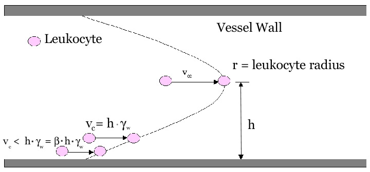
In intravital microscopy experiments, the velocity of a cell flowing in the centerline (vcc) can be determined experimentally. From vcc, the mean blood flow velocity, vB, can be calculated as
Wall shear rate in microvessels is estimated as
where d is the vessel diameter and 2.12 is an empirical correction factor based on velocity profiles measured in vivo (Reneman, 1992). From gw, critical velocity is calculated as
where b is a dimensionless drag coefficient derived from theoretical analysis of a sphere flowing close to a wall (Goldman, 1967). Values of b are listed in a table below. As an approximation, for a clearance (h - r) of about 50 nm, the critical velocity can be estimated as
where e is the ratio of the leukocyte diameter (assumed to be 7mm) to the vessel diameter. Any cell traveling at a velocity below the critical velocity is considered a rolling cell, as it is likely to be retarded by an adhesive interaction with the vessel wall.
|
|
Absolute clearance of cell |
b |
|
|
|
|
|
|
|
|
|
|
|
|
|
|
|
|
|
|
|
|
|
|
|
|
|
|
|
|
|
|
|
|
|
|
|
|
| Read the following categorized abstracts or link to Medline to conduct your own query.
|
Comment on this page's content or view comments made by others!